|
|
|
|
BABCERC Scientific Publications
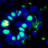
Article summary Background:Exposure to radiation is known to increase breast cancer risk. It is also known that this risk is dose-dependent, which means the more exposure that occurs, the greater the cancer risk. It has long been believed that radiation causes cancer by damaging a cell’s DNA. More recently, though, researchers have realized that radiation exposure also can induce molecular signals, like cytokines, that cells use to communicate with one another. These communication problems can affect the environment that surrounds a cell. They also can cause a cell to change in ways that make it more likely to become a cancer cell. Such changes are referred to as “phenotype” because even though the cell has changed, its DNA has remained the same. Cell phenotype is established by changes in how the DNA marks, which are called “epigenetic”. Just as the human body consists of 300 cells types that remain ‘true’ throughout life, studies have shown that when a cell that has developed a pre-cancerous phenotype, it can pass on these change to its daughter cells. It’s currently not known whether and to what extent this type of heritable cell behaviors contribute to the development of breast or other cancers. However it has been shown that when placed into a laboratory dish cells from a normal human breast consist of two phenotypes: most live just a short time before they are unable to divide any longer but another rare phenotype can live weeks longer. It is not known why variations in cell phenotype are present in normal breast but it is thought that the long-lived ones have a greater potential to turn into cancer cells because a protein called p16 is suppressed by epigenetic markings, called methylation. But it is known that human mammary cells only grow if there is room for the daughter cells to spread out. So if the dish is full, the cells stop growing. Study Design: The research team performed this study in a laboratory using breast epithelial cells that had been taken from normal breast tissue. (The epithelial cells are the cells that line the breast duct, which is where most breast cancers begin.) The cells were grown in the laboratory for seven to eight days then the researchers exposed the cells to a single low-to-moderate dose of radiation. The researchers also grew breast cells that they did not irradiate, which allowed them to compare differences between exposed and unexposed cells. The research team then evaluated whether radiation affected the growth of cells. Results:
Conclusions:In this study, the researchers showed that radiation could indirectly promote the growth of long-lived variant cells by speeding up the aging process of normal cells. By getting normal cells to prematurely age and stop dividing, radiation exposure can create a space for epigenetically altered cells to fill the void these cells would have normally filled. These findings support the hypothesis that radiation can cause cancer by making the environment that surrounds pre-cancerous cells more hospitable to tumor growth. This suggests that to fully understand how radiation causes cancer, researchers must study the effect radiation has on a cell’s microenvironment. This study was jointly funded the National Institute of Environmental Health Sciences (NIEHS), and the National Cancer Institute (NCI), NIH, through the Bay Area Breast Cancer and the Environment Center and by NASA Specialized Center of Research.Article citation: Address correspondence to Dr. Paul Yaswen. Life Sciences Division, Lawrence Berkeley National Laboratory, 1 Cyclotron Road, Berkeley, CA 94720. E-mail: [email protected] Link to article: http://breast-cancer-research.com/content/12/1/R11 |
Funded by the National Institute of Environmental Health Sciences (NIEHS) and the National Cancer Institute (NCI), NIH, DHHS. |
|